
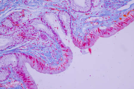
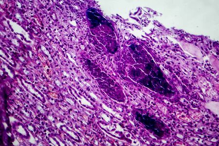
Condyloma acuminatum, also known as genital warts. Light micrograph, photo under microscope
Коллекция по умолчанию
Коллекция по умолчанию
Создать новую

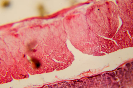
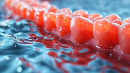
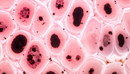
Abstract science background- pyloric division of the stomach of the dog. Cell biology
Коллекция по умолчанию
Коллекция по умолчанию
Создать новую

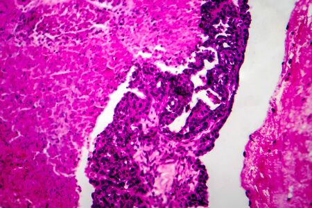
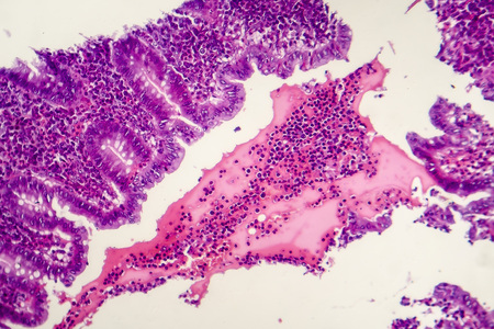
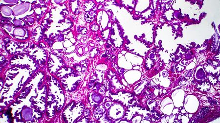
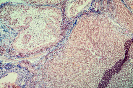
Ovarian cancer, light micrograph, photo under microscope. Photograph shows a fragment of a cancerous tumor in the female ovary. Selective focus
Коллекция по умолчанию
Коллекция по умолчанию
Создать новую
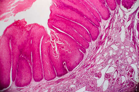
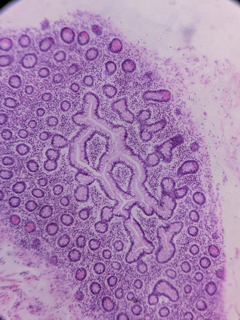
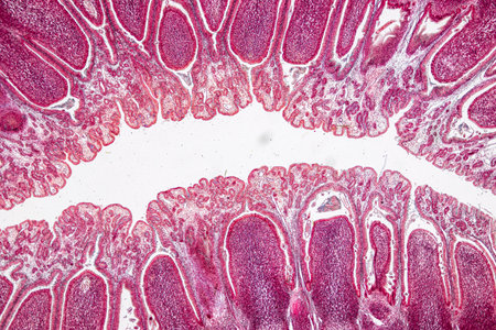
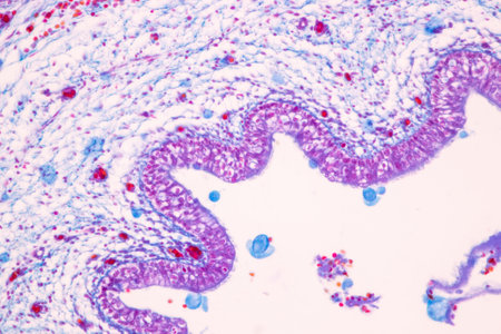
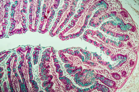
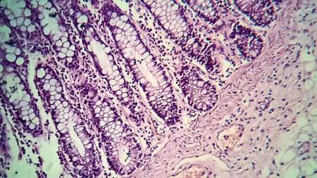
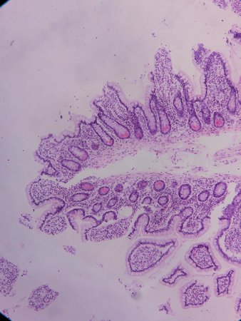
Intestinal wall section showing villi and epithelial lining clearly
Коллекция по умолчанию
Коллекция по умолчанию
Создать новую

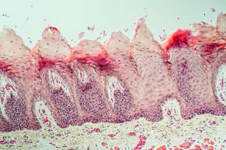
Cross section of human skin under microscope view for education in laboratory.
Коллекция по умолчанию
Коллекция по умолчанию
Создать новую
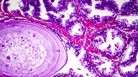
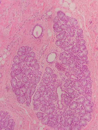
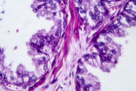
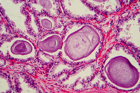
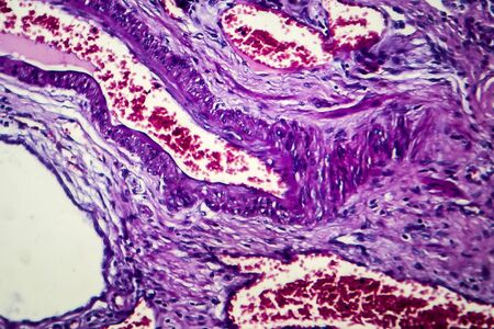
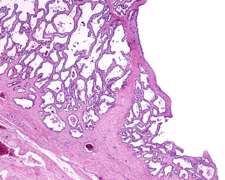
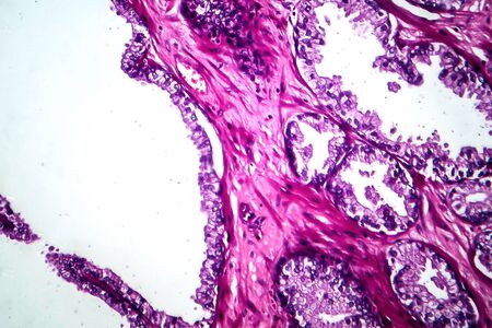
Photomicrograph showing histological features of benign prostatic hyperplasia. Enlarged prostate gland with nodular proliferation of glandular and stromal components.
Коллекция по умолчанию
Коллекция по умолчанию
Создать новую
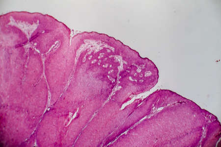
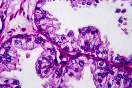
Condyloma acuminatum, also known as genital warts. Light micrograph, photo under microscope
Коллекция по умолчанию
Коллекция по умолчанию
Создать новую
Ice texture background, ink in water pattern frost. Crystal winter design
Коллекция по умолчанию
Коллекция по умолчанию
Создать новую

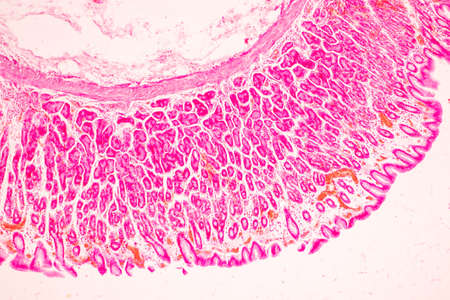
Stomach tissue under the microscope 100x
Коллекция по умолчанию
Коллекция по умолчанию
Создать новую
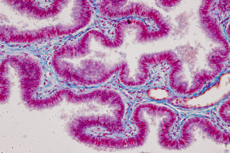
Tissue of Stomach Human under the microscope in Lab.
Коллекция по умолчанию
Коллекция по умолчанию
Создать новую
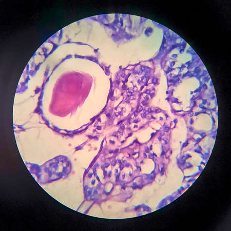
Pathology and Histology Tissue of Mammals under microscope.
Коллекция по умолчанию
Коллекция по умолчанию
Создать новую
Thyroid follicular carcinoma, light micrograph, photo under microscope
Коллекция по умолчанию
Коллекция по умолчанию
Создать новую
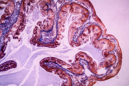
Columnar epithelium of human gall bladder under the microscope in Lab.
Коллекция по умолчанию
Коллекция по умолчанию
Создать новую

Histological Uterus human, Uterine tube human, Placenta human and Umbilical cord Human under the microscope for education.
Коллекция по умолчанию
Коллекция по умолчанию
Создать новую
Showing Light micrograph of the Thyroid gland and Thymus gland human Child under the microscope for education in the laboratory.
Коллекция по умолчанию
Коллекция по умолчанию
Создать новую
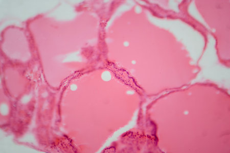
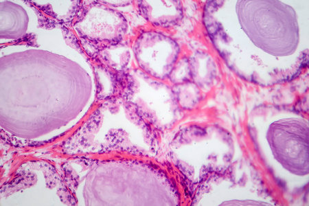
Photomicrograph of a normal thyroid gland under a microscope, exhibiting typical follicular structure and colloid-filled follicles.
Коллекция по умолчанию
Коллекция по умолчанию
Создать новую

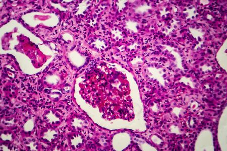
Histopathology of acute nephritis, light micrograph, photo under microscope
Коллекция по умолчанию
Коллекция по умолчанию
Создать новую
Histopathology of prostate gland hyperplasia, light micrograph, photo under microscope
Коллекция по умолчанию
Коллекция по умолчанию
Создать новую
Pancreas cancer cell under microscope view for medical education.
Коллекция по умолчанию
Коллекция по умолчанию
Создать новую
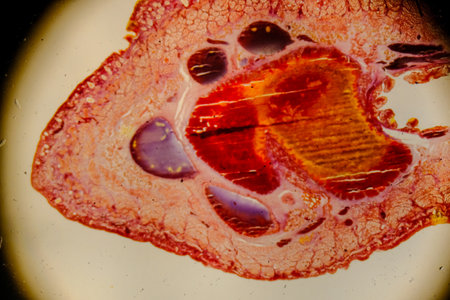
Leech cross section showing internal anatomical structures stained
Коллекция по умолчанию
Коллекция по умолчанию
Создать новую
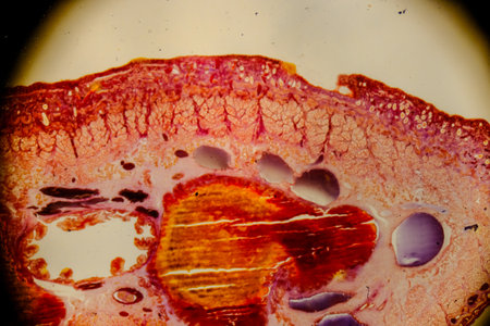
Leech cross section showing internal anatomical structures stained
Коллекция по умолчанию
Коллекция по умолчанию
Создать новую

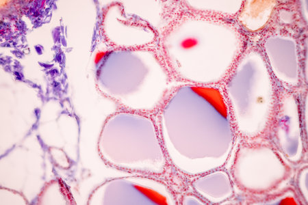
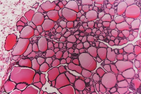
Endemic goiter, light micrograph, abnormal enlargement of the thyroid gland due to dietary iodine deficiency. Photomicrograph shows follicles of varying size, abundant colloid, lymphocytic infiltrate
Коллекция по умолчанию
Коллекция по умолчанию
Создать новую
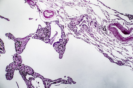
Histopathology of lung emphysema, light micrograph, photo under microscope showing enlargement of air spaces in lung tissue and destruction of alveolar septa
Коллекция по умолчанию
Коллекция по умолчанию
Создать новую
Pathology and Histology Tissue of Mammals under microscope.
Коллекция по умолчанию
Коллекция по умолчанию
Создать новую
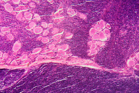
A longitudinal section of human spinal ganglion cells under the microscope.
Коллекция по умолчанию
Коллекция по умолчанию
Создать новую
Stomach tissue under the microscope 100x
Коллекция по умолчанию
Коллекция по умолчанию
Создать новую

Endometriosis, a disorder in which cells similar to those in the endometrium grow outside the uterus. Light micrograph, photo under microscope
Коллекция по умолчанию
Коллекция по умолчанию
Создать новую
Photomicrograph showing histological features of benign prostatic hyperplasia. Enlarged prostate gland with nodular proliferation of glandular and stromal components.
Коллекция по умолчанию
Коллекция по умолчанию
Создать новую
Acute pyelonephritis, light micrograph, photo under microscope
Коллекция по умолчанию
Коллекция по умолчанию
Создать новую
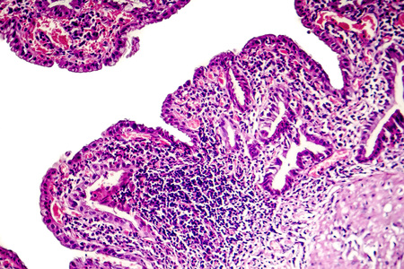
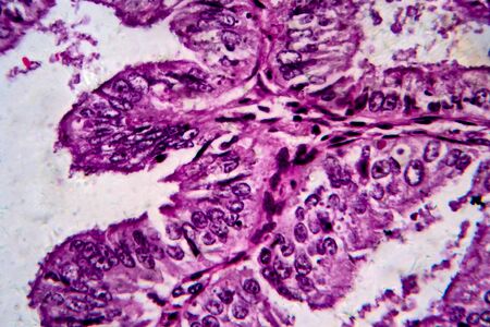
Papillary serous ovarian adenocarcinoma, cancer of ovary, light micrograph, photo under microscope
Коллекция по умолчанию
Коллекция по умолчанию
Создать новую
dentistry, concept.
Коллекция по умолчанию
Коллекция по умолчанию
Создать новую
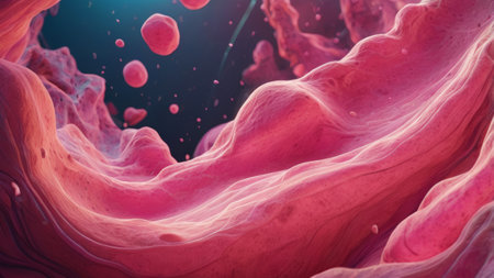
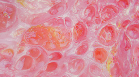
Macro view of colorful abstract texture, resembling organic biological forms. Bright pink and red hues create a dynamic and fluid pattern.
Коллекция по умолчанию
Коллекция по умолчанию
Создать новую
Showing Light micrograph of the Trachea, Thymus, Parathyroid gland and Tonsil human under the microscope for education in the laboratory.
Коллекция по умолчанию
Коллекция по умолчанию
Создать новую
Histopathology of human under microscope view for education in laboratory.
Коллекция по умолчанию
Коллекция по умолчанию
Создать новую
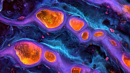
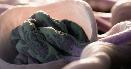
See vibrant neurons ignite during creative challenges! Stunning visualizations reveal enhanced brain function, showcasing improved focus, concentration, and problem-solving Witness the beauty of neurological processes under pressure as cognitive skills are amplified AI Generative
Коллекция по умолчанию
Коллекция по умолчанию
Создать новую
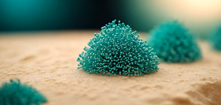
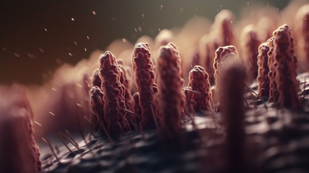
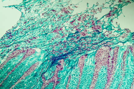
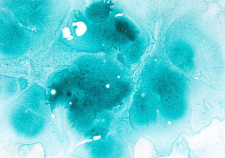
Explore a fascinating, alien world with this striking microscopic view of vibrant turquoise flora on a textured sandy surface, perfect for science and sci-fi concepts.
Коллекция по умолчанию
Коллекция по умолчанию
Создать новую
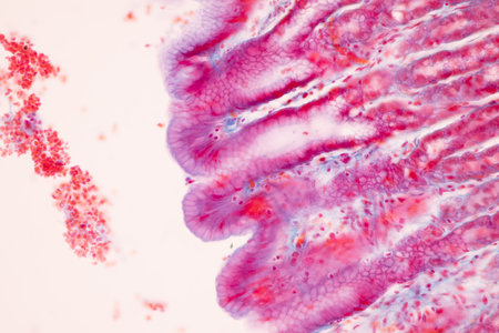
Small intestine with villi under the microscope 100x
Коллекция по умолчанию
Коллекция по умолчанию
Создать новую

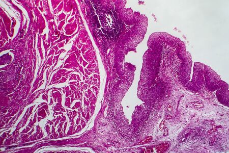
Colon polyp, one of the largest polyps
Коллекция по умолчанию
Коллекция по умолчанию
Создать новую
A microscopic view of tissue with pink and purple staining, showing cellular structures and patterns
Коллекция по умолчанию
Коллекция по умолчанию
Создать новую
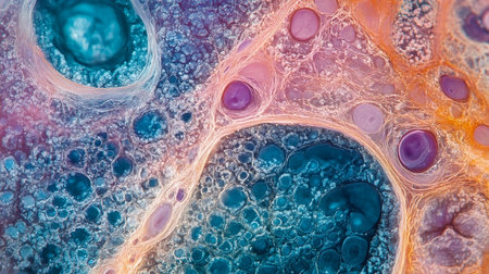
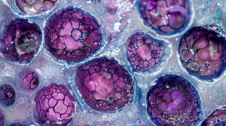
A stunning close-up of colorful cells, showcasing intricate patterns and textures. The vibrant colors and details illustrate the beauty of microscopic biology in an abstract way. Generative AI
Коллекция по умолчанию
Коллекция по умолчанию
Создать новую

Histopathology of lung emphysema, light micrograph, photo under microscope showing enlargement of air spaces in lung tissue and destruction of alveolar septa
Коллекция по умолчанию
Коллекция по умолчанию
Создать новую
Human liver tissue under microscope view. Histological sample of human liver.
Коллекция по умолчанию
Коллекция по умолчанию
Создать новую

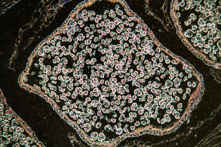
Reproductive parts: stigma, style, stamens, filament, petal. Structure floral- male reproductive organ of the flower of angiosperms
Коллекция по умолчанию
Коллекция по умолчанию
Создать новую
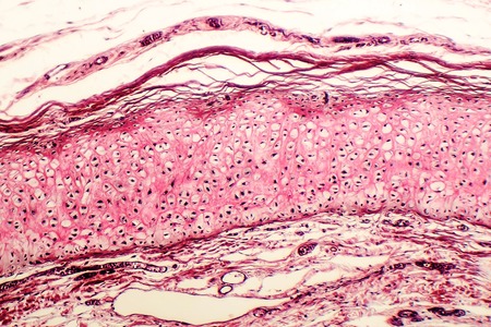
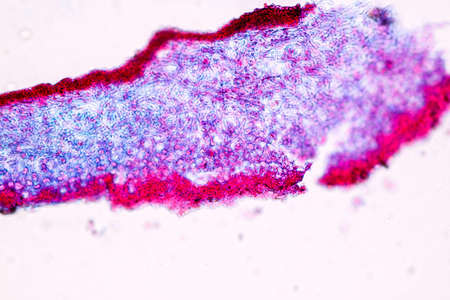
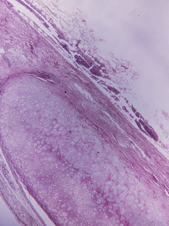
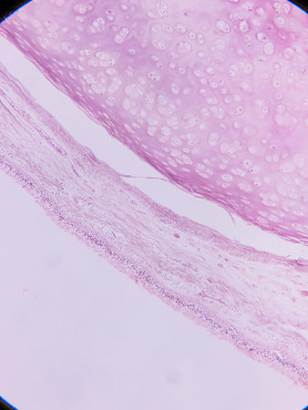
Elastic cartilage of human outer ear, light micrograph
Коллекция по умолчанию
Коллекция по умолчанию
Создать новую
Bacillary dysentery, light micrograph, photo under microscope showing presence of bacteria and accumulation of inflammatory cells in intestinal epithelium
Коллекция по умолчанию
Коллекция по умолчанию
Создать новую
Abstract science- biology tissue thyroid of dog. Medical and biological tissue prepared microscope slide. Educational material for the study and treatment of animals.
Коллекция по умолчанию
Коллекция по умолчанию
Создать новую
Macro shot of microbes cells and bacteria, 3d microscopic virus close up, abstract background or fantasy wallpaper, by ai generative
Коллекция по умолчанию
Коллекция по умолчанию
Создать новую

Columnar epithelium of human gall bladder under the microscope in Lab.
Коллекция по умолчанию
Коллекция по умолчанию
Создать новую
macro image of many white blood cells created using AI
Коллекция по умолчанию
Коллекция по умолчанию
Создать новую
Histopathology of cholera under microscope view for education.
Коллекция по умолчанию
Коллекция по умолчанию
Создать новую
Club moss flower with seeds under the microscope 100x
Коллекция по умолчанию
Коллекция по умолчанию
Создать новую
Characteristics of Lichen, hyphae and Symbiotic algae under the microscope for education.
Коллекция по умолчанию
Коллекция по умолчанию
Создать новую
Prostate cancer, light micrograph, photo under microscope
Коллекция по умолчанию
Коллекция по умолчанию
Создать новую

Endometriosis, a disorder in which cells similar to those in the endometrium grow outside the uterus. Light micrograph, photo under microscope
Коллекция по умолчанию
Коллекция по умолчанию
Создать новую
Tongue Tissue with taste buds across 200x
Коллекция по умолчанию
Коллекция по умолчанию
Создать новую
Prostate cancer, light micrograph, photo under microscope
Коллекция по умолчанию
Коллекция по умолчанию
Создать новую

Chronic nephritis, light micrograph, photo under microscope
Коллекция по умолчанию
Коллекция по умолчанию
Создать новую
Photomicrograph showing histological features of benign prostatic hyperplasia. Enlarged prostate gland with nodular proliferation of glandular and stromal components.
Коллекция по умолчанию
Коллекция по умолчанию
Создать новую
Chronic nephritis, light micrograph, photo under microscope
Коллекция по умолчанию
Коллекция по умолчанию
Создать новую

Cross section of human body under microscope view for education in laboratory.
Коллекция по умолчанию
Коллекция по умолчанию
Создать новую
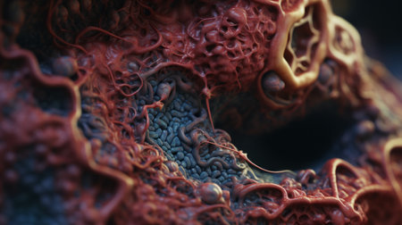
a close-up photo of a highly detailed organ, resembling a zbrush creation. the organ showcases vibrant red cells, while the intricate anatomy is captured with the zeiss batis 18mm f2.8 lens. rendered in maya, the photo features a unique squiggly line style, with texture-rich compositions and mesmerizing swirling vortexes. ai generated
Коллекция по умолчанию
Коллекция по умолчанию
Создать новую
Vibrant strawberry rhubarb jam texture with abstract patterns for food design
Коллекция по умолчанию
Коллекция по умолчанию
Создать новую
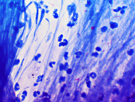
Mycobacterium tuberculosis positive (small red rod) in sputum smear, acid-fast stain, analyze by microscope 1000x
Коллекция по умолчанию
Коллекция по умолчанию
Создать новую
A stunning microscopic view captures the intricate dance of life as plant cells undergo mitosis. Each stained cell reveals a different stage of division, illustrating the fundamental process of growth and reproduction in a vibrant, detailed display.
Коллекция по умолчанию
Коллекция по умолчанию
Создать новую
Colon cancer tissue under the microscope 100x
Коллекция по умолчанию
Коллекция по умолчанию
Создать новую
Suppurative appendicitis, light micrograph, photo under microscope showing neutrophilic infiltrates of the appendix wall and lumen
Коллекция по умолчанию
Коллекция по умолчанию
Создать новую
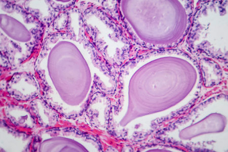
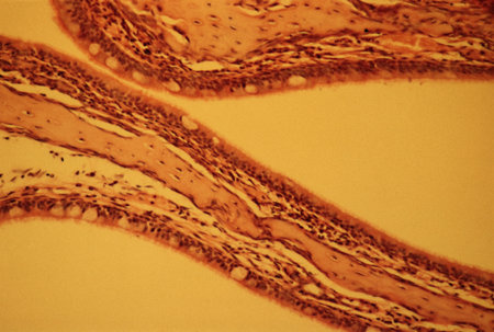
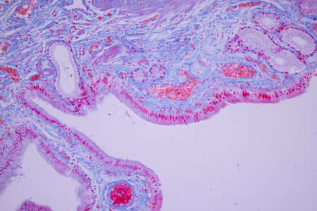
Human seminal vesicle. The surface of the mucosa is very folded. The spaces that look like glands are really infoldings of the mucosa that communicate with the lumen. The epithelium is pseudostratified columnar with basal cells.
Коллекция по умолчанию
Коллекция по умолчанию
Создать новую

Education anatomy and Histological sample Spinal cord Tissue under the microscope.
Коллекция по умолчанию
Коллекция по умолчанию
Создать новую
Prostate cancer, light micrograph, photo under microscope
Коллекция по умолчанию
Коллекция по умолчанию
Создать новую

Histological Spermatic cord human, Seminal vesicle human, Prostate human and Human chromosomes under the microscope for education.
Коллекция по умолчанию
Коллекция по умолчанию
Создать новую
Histopathology of prostate gland hyperplasia, light micrograph, photo under microscope
Коллекция по умолчанию
Коллекция по умолчанию
Создать новую
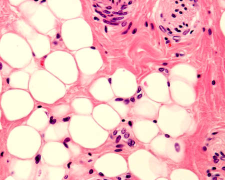
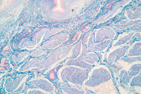
Small adipocyte lobule located in a connective tissue. A small nerve is located in the upper right corner. Light micrograph. H&E stain.
Коллекция по умолчанию
Коллекция по умолчанию
Создать новую
Closeup of structure of microbiotic flora of human body. Microscopic bacteria cells. Science concept. Generative AI.
Коллекция по умолчанию
Коллекция по умолчанию
Создать новую
Coccidiosis of liver tissue under the microscope 100x
Коллекция по умолчанию
Коллекция по умолчанию
Создать новую
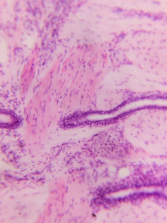
Microscopic view of tissue section showing cellular structures and layers, stained for examination
Коллекция по умолчанию
Коллекция по умолчанию
Создать новую
Painting acrylic paint- abstract drawing. Texture background
Коллекция по умолчанию
Коллекция по умолчанию
Создать новую
Chronic cholecystitis, light micrograph, photo under microscope showing fibrosis and muscular hypertrophy of gallbladder wall, entrapped epithelial crypts, foamy macrophages
Коллекция по умолчанию
Коллекция по умолчанию
Создать новую
Endometrial adenocarcinoma, light micrograph, photo under microscope
Коллекция по умолчанию
Коллекция по умолчанию
Создать новую
Education anatomy and Histological sample of Human under the microscope.
Коллекция по умолчанию
Коллекция по умолчанию
Создать новую
Nasal cavity
Коллекция по умолчанию
Коллекция по умолчанию
Создать новую
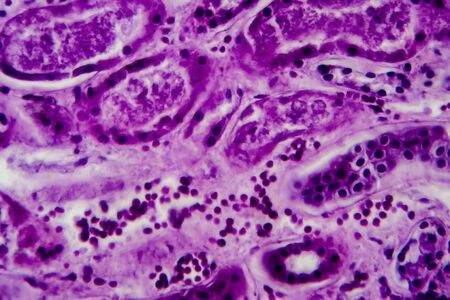
Diffuse proliferative glomerulonephritis, light micrograph, photo under microscope. High magnification
Коллекция по умолчанию
Коллекция по умолчанию
Создать новую
Histopathology of human under microscope view for education in laboratory.
Коллекция по умолчанию
Коллекция по умолчанию
Создать новую
Columnar epithelium of human gall bladder under the microscope in Lab.
Коллекция по умолчанию
Коллекция по умолчанию
Создать новую

Ice texture background, ink in water pattern frost. Crystal winter design
Коллекция по умолчанию
Коллекция по умолчанию
Создать новую

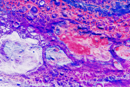
Bladder cat- cell nature background. Abstract- photo macro sections with high magnification with light microscope
Коллекция по умолчанию
Коллекция по умолчанию
Создать новую
Esophageal squamous cell carcinoma, light micrograph, photo under microscope
Коллекция по умолчанию
Коллекция по умолчанию
Создать новую

Characteristics of Lichen, hyphae and Symbiotic algae under the microscope for education.
Коллекция по умолчанию
Коллекция по умолчанию
Создать новую
Showing Light micrograph of the Adrenal gland and Urinary bladder human under the microscope for education in the laboratory.
Коллекция по умолчанию
Коллекция по умолчанию
Создать новую
Chronic pyelonephritis, light micrograph, photo under microscope
Коллекция по умолчанию
Коллекция по умолчанию
Создать новую

Tissue of Small intestine (Duodenum) and Vermiform appendix Human under the microscope in Lab.
Коллекция по умолчанию
Коллекция по умолчанию
Создать новую
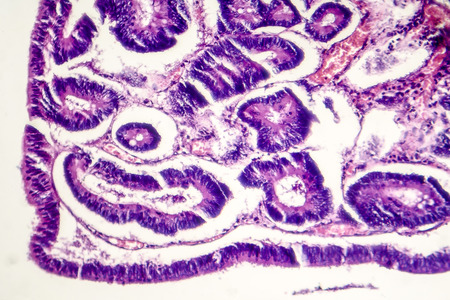
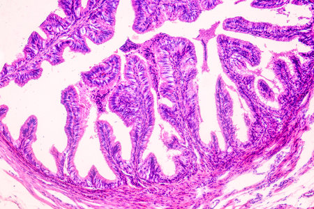
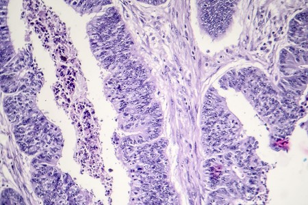
Intestinal polypoid adenoma, light micrograph, photo under microscope
Коллекция по умолчанию
Коллекция по умолчанию
Создать новую
Extreme Close up of microscopic kidney Bowman's Capsule and Glomerulus
Коллекция по умолчанию
Коллекция по умолчанию
Создать новую

Kidney cancer, light micrograph, photo under microscope. High magnification
Коллекция по умолчанию
Коллекция по умолчанию
Создать новую
blue paint Abstract art background texture watercolor on paper Modern design of posters, cards, invitations, wallpapers
Коллекция по умолчанию
Коллекция по умолчанию
Создать новую

Nature pattern: cross-section weevil rye. Beautiful vegetable background. Biological prepared microscope slides
Коллекция по умолчанию
Коллекция по умолчанию
Создать новую
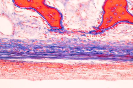
Anatomy and Histological Bone, Elastic cartilage human and Joint of human foetus under the microscope for education.
Коллекция по умолчанию
Коллекция по умолчанию
Создать новую

Cell- science background. Esophagus of the dog- cross section
Коллекция по умолчанию
Коллекция по умолчанию
Создать новую
Bladder cancer, light micrograph, photo under microscope
Коллекция по умолчанию
Коллекция по умолчанию
Создать новую
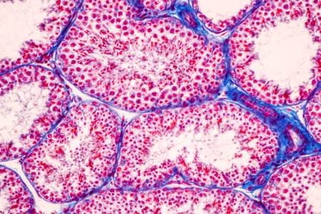
Anatomy and Histological Epididymis and Testis human cells under microscope.
Коллекция по умолчанию
Коллекция по умолчанию
Создать новую
Microscopic view of the bone marrow stroma shows colonies of mesenchymal stem cells which have the ability to differentiate into various cell types
Коллекция по умолчанию
Коллекция по умолчанию
Создать новую
Legion-Media
Создайте свои проекты на основе качественных стоковых фотографий и видео.


Copyright © Legion-Media.